electric blanket pad
-
2. Ease of Use Electric heating mats are incredibly user-friendly. They usually come with adjustable temperature settings, allowing users to customize their heating experience based on personal preferences. Many models feature timers and auto shut-off functions for added safety, ensuring that the mats turn off when not in use.
electric heating mat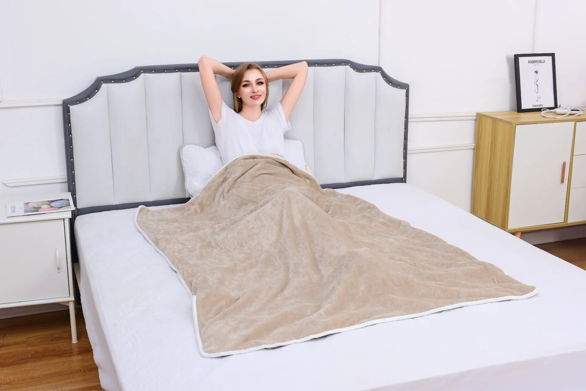
...
Top Ranking
-
1
As the chill of winter sets in, many individuals seek effective ways to stay warm without significantly increasing their energy bills. Low energy heated blankets have emerged as a practical solution, offering comfort and warmth while being mindful of energy consumption. These blankets are not just a luxury; they are an investment in both comfort and eco-friendliness, making them an ideal addition to any household.
-
2
In summary, twin heated blankets are an excellent addition to any home during the colder months. They offer unparalleled comfort, energy efficiency, and versatility, making them ideal for a wide range of situations. With safety features and easy maintenance, a twin heated blanket is not just a luxury but a practical investment in your comfort and well-being. As winter approaches, consider adding one to your bedding ensemble and enjoy the cozy warmth it brings on those chilly nights.
-
3
In conclusion, the cost of calming heat back wraps can vary greatly depending on several factors including material quality, technological features, brand reputation, and additional functionalities. Potential buyers are encouraged to assess their specific needs and preferences when choosing a wrap. While investing in a quality calming heat back wrap may require a larger upfront expense, the potential benefits it brings in terms of pain relief and stress management may justify the investment, contributing to a more comfortable and relaxing lifestyle. As with any wellness product, it’s essential to do thorough research and choose a model that best aligns with individual needs and budget considerations.
-
4
The Comfortable Embrace of Electric Throw Blankets A Double Delight
-
5
The Comfort of Sleeping with a Heated Blanket
-
6
Safety Features
-
7
4. Pain Relief For individuals suffering from chronic pain, such as arthritis or muscle soreness, the gentle heat of a heated blanket can offer therapeutic benefits. The heat can help alleviate tension in muscles, reduce pain, and promote relaxation, making them an excellent addition to any wellness routine.
heated blanket cord
-
'> 8Safety is a common concern when it comes to electric blankets, but modern designs come equipped with advanced safety features. Many electric blankets now include an automatic shut-off function that turns the blanket off after a certain period, reducing the risk of overheating or electrical malfunctions. Furthermore, they often feature flexible wires and low-voltage technology, making them safer than older models. It is essential for consumers to follow the manufacturer's instructions and guidelines to ensure safe use.
Netizens pay attention
-
Heat therapy is particularly effective in relaxing tense and tight muscles. After a long day at the office or an intense workout, the muscles in your back may become rigid and sore. A heating pad helps to soften these tight spots, allowing for greater flexibility and reducing the risk of further injury. By incorporating heat therapy into your routine, you can significantly enhance your overall mobility and well-being.
heating pad for whole back
2025-08-13 23:34Read1937Visitors
Looked and looked
- Material Look for a bed warmer made from soft, breathable fabrics to ensure comfort against your skin. Some options are designed with hypoallergenic materials, perfect for those with sensitivities.
- Materials Look for soft, high-quality materials that feel good against the skin, such as fleece or micro plush. These materials not only enhance comfort but also retain heat effectively.
Using a soft heat heating pad is also an effective way to manage stress. Heat has a calming effect on the body, which can help to reduce stress levels and promote relaxation. After a long day, spending some time with a heating pad can serve as a simple yet effective self-care ritual that enhances the overall quality of life.
soft heat heating pad
'>A double bed hot blanket is designed specifically for dual occupancy, ensuring that both sides of the bed are equally warm and inviting. Unlike traditional blankets that rely solely on your body heat, a hot blanket is equipped with heating elements that evenly distribute warmth across its surface. This provides an immediate, consistent comfort that traditional blankets simply can't match.
The Comfort of Electric Blankets for Over the Knees
'>In addition to physical benefits, using a heat pad for your feet in bed can also have psychological advantages. The sensation of warmth can trigger the release of endorphins, which are natural mood lifters. The soothing heat can reduce levels of cortisol, the stress hormone, leading to an overall feeling of relaxation and well-being. When you're less stressed, you're more likely to have a peaceful night's sleep, further enhancing your mental clarity and emotional resilience during the day.
2. Muscle Relaxation For athletes or those who engage in physical activity, muscle tightness is commonplace. Applying heat to the muscles can help them relax and release tension, thereby reducing the risk of injury. By using a body length heating pad after a workout, you can prevent soreness and promote faster recovery.
body length heating pad
- Washability Having an easily washable electric blanket is vital for hygiene, especially since dogs may track in dirt and hair. Look for blankets with removable covers that can be machine washed.